Posts

Clash of Neurological Titans: Guillain-Barré Syndrome vs Acute Transverse Myelitis
Image
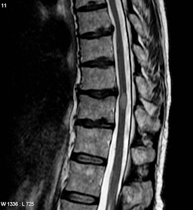
Guillain-Barré Syndrome vs Acute Transverse Myelitis  Both Transverse myelitis and Guillain-Barre syndrome are immunologically caused polyneuropathies with significant clinical implications. Although no precise genetic risk loci have been identified as of yet, both are believed to have a hereditary tendency. Both are regarded as autoimmune diseases, but the exact causes are not yet known. Both may be brought about by molecular mimicry, especially from vaccinations and infectious agents, but it is obvious that host factors and co-founding host responses will affect the natural history  and susceptibility to disease. The symptoms of GBS, an acute inflammatory immune-mediated polyradiculoneuropathy, include discomfort, tingling, and increasing weakness as well as autonomic dysfunction. In acute inflammatory demyelinating polyneuropathy, immune destruction occurs specifically at the myelin sheath and associated Schwann-cell components; in acute motor axonal neuropathy, however, th...

Decoding Nothnagel Syndrome: Navigating the Mysteries of Rare Neurological Disorder

Image
 Nothnagel Syndrome        The clinical characteristics of malignancies involving the quadrigeminal bodies were outlined by Austrian physician Nothnagel in his monograph, Localization of Diseases of the Brain (1879). He limited his talk to neoplasms since he believed they were more specific to this area than cerebrovascular infarctions.  Hermann Nothnagel presented a 2 year old child who had dilated pupils, significant gait ataxia, and a vague bilateral ophthalmoparesis. After an autopsy, it was discovered that the patient had hydrocephalus and a sarcoma that had invaded all 4 colliculi, the pineal gland, and the cerebellum dorsally. Nothnagel came to several conclusions following a through examination of the literature 1) tumors involving the inferior colliculi can cause bilateral oculomotor paralysis of varying degrees in each eye, and 2) lesions of the superior colliculi may be linked to abnormal pupillary responses and visual loss without papilledema or...

Spinal Showdown: Cauda Equina Syndrome vs. Conus Medullaris Syndrome

Image
Cauda Equina Syndrome vs. Conus Medullaris Syndrome CAUDA EQUINA SYNDROME: Anatomy The term "cauda equina" refers to the group of nerves at the end of the spinal cord that resembles the tail of a horse. At the top of the lumbar (lower back) spine, the spinal cord terminates. The spinal canal contains the separate nerve roots that continue from the spinal cord's end and give the legs and bladder sensory and motor function. Following these nerve roots into the lumbar and sacral regions is the cauda equina. The lower limbs and pelvic organs are communicated with by these nerves both inside and externally.    Spinal nerves L2-L5, S1-S5, and Co1 continue inferiorly as the cauda equina. Compression of these nerves can produce cauda equina or conus medullaris syndromes.  Cauda equina syndrome (CES) occurs when there is dysfunction of multiple lumbar and sacral nerve roots of the cauda equina.  Etiology Spinal lesions and tumors Lumbar spinal stenosis Birth abnormalities...

Unlocking the Mysteries of Lateral Medullary Syndrome: A Journey through Brainstem Disruption

Image
Lateral Medullary Syndrome, also known as Wallenberg Syndrome Clinical Picture : A 65 M presents with sudden onset ataxia, slurred speech and vomiting. Known case of Hypertension  and Diabetes mellitus . On examination he has no power loss in the limbs, features of Horner syndrome on right side and loss of pain and temperature sensation on right side of the body and left side of his face. Other systems are normal. (Vishram Singh neuroanatomy) The region infarcted in lateral medullary syndrome is seen in the illustration above. The posterior inferior cerebellar artery, a branch of the vertebral artery, is the artery that gets injured.  (Snells Neuroanatomy) With the aid of the simple neuroanatomy of the medulla and its section at the level of the inferior olivary nucleus, as previously demonstrated, we may determine which structures are affected. Structures involved are: Inferior Cerebellar Peduncle  Vagus nerve nuclei - Nucleus tractus solit...

Unraveling the Marvels of the Mind: Your Ultimate Brain Blog!

Image
Hello,  I am Shardool Gadgil, a medical student from Mumbai, India. This blog is devoted to discussing the amazing and complicated brain and how its complex structure may be used to better understand the numerous illnesses and disorders that affect the nervous system. Gaining knowledge of neuroanatomy creates new avenues for comprehending and responding to fundamental questions in the medical sciences, such as where the lesion is, how it happens, why these symptoms and signs appear, and what the disease's pathophysiology is.  This will be a weekly blog most probably with links to various resources available to understand the neurosciences.   Let us explore the enormous reservoir of information our brains can hold and find the answers to some of the most fascinating queries we encounter.  Let's eliminate the NEUROPHOBIA!!!