Clash of Neurological Titans: Guillain-Barré Syndrome vs Acute Transverse Myelitis
Guillain-Barré Syndrome vs Acute Transverse Myelitis

Both Transverse myelitis and Guillain-Barre syndrome are immunologically caused polyneuropathies with significant clinical implications. Although no precise genetic risk loci have been identified as of yet, both are believed to have a hereditary tendency. Both are regarded as autoimmune diseases, but the exact causes are not yet known. Both may be brought about by molecular mimicry, especially from vaccinations and infectious agents, but it is obvious that host factors and co-founding host responses will affect the natural history and susceptibility to disease.
The symptoms of GBS, an acute inflammatory immune-mediated polyradiculoneuropathy, include discomfort, tingling, and increasing weakness as well as autonomic dysfunction. In acute inflammatory demyelinating polyneuropathy, immune destruction occurs specifically at the myelin sheath and associated Schwann-cell components; in acute motor axonal neuropathy, however, the primary target for immune-related harm is the membranes on the nerve axon, or the axolemma.
Patients with GBS related to infections frequently produce antibodies against human peripheral nerve gangliosides
On the other hand, acute or subacute motor, sensory, and autonomic spinal cord dysfunction is a hallmark of TM, an inflammatory condition. Similar to GBS, there is disruption of ascending and descending neuroanatomical circuits in the transverse spinal cord plane. Molecular mimicry and infectious organisms have been proposed as triggers.
Transverse Myelitis
The clinical signs of TM are caused by neurological dysfunction of motor, sensory, and autonomic pathways within and passing through the inflamed area. TM is defined by focal inflammation within the spinal cord. A spinal MRI and lumbar puncture frequently reveal a well-defined rostral border of sensory impairment as well as acute inflammation.
Diagnostic criteria for Transverse Myelitis proposed by the Transverse Myelitis Consortium Working Group:
| Inclusion criteria for diagnosis of transverse myelitis (idiopathic or disease associated) | |
| Development of sensory, motor or autonomic dysfunction attributable to the spinal cord | |
| Bilateral symptoms | |
| Clearly defined sensory level | |
| Exclusion of compressive aetiology by MRI or CT myelography | |
| Spinal cord inflammation demonstrated by CSF pleocytosis or elevated IgG or gadolinium enhancement | |
| Progression to clinical nadir between 4 hours and 21 days from onset of symptoms | |
| Exclusion criteria for diagnosis of transverse myelitis (idiopathic or disease associated) | |
| History of radiation to the spine within 10 years | |
| Clear arterial distribution clinical defect consistent with anterior spinal artery occlusion | |
| Abnormal flow voids on the surface of the cord consistent with AVM | |
| Exclusion criteria for idiopathic transverse myelitis | |
| Serologic or clinical evidence of connective tissue disease (sarcoidosis, Behcet's disease, Sjogren's syndrome, SLE, mixed connective tissue disorder) | |
| CNS manifestations of syphilis, Lyme disease, HIV, HTLV-1, Mycoplasma, other viral infection. | |
| Brain abnormalities suggestive of MS | |
| History of clinically apparent optic neuritis | |

There are a number of inflammatory conditions that appear to cause the disorder:
- Multiple Sclerosis
- Sarcoidosis
- Neuromyelitis optica (Devic's disease)
- Vaccinations
- It is likely that some individuals with transverse myelitis have autoimmune diseases. These conditions include Sjogren's syndrome, which results in very dry mouth and eyes, and lupus, which can impact several bodily systems.
A useful article about Transverse Myelitis.
Neuroanatomy Magic:


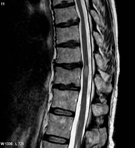
(MRI spine showing a hyperdense white cord lesion)
Understanding the neuroanatomy of the spinal cord goes hand in hand in understanding the patient presentation.
Depending on the the level of cord affected the signs and symptoms differ, for example if the cervical cord is affected the patient might become quadriplegic, if the thoracic or lumbar cord is affected that will manifest as paraplegia and if the sacral cord is affected that might show associated bladder symptoms.
Understanding the anatomy of the tracts in the spinal cord is very important to diagnose transverse myelitis. In the first image above showing the transverse section of spinal cord with the neuroanatomical placement of various ascending and descending tracts:
Ascending tracts:
- Anterior and lateral spinothalamic tracts: These tracts carry the sensations like pain, touch and temperature from the peripheral receptors to the cortex. The first order neurons enter the cord through the dorsal root ganglion and relay in the cord. The second order neurons cross immediately to the opposite side of the spinal cord and ascend up the cord as the anterior and lateral spinothalamic tracts.
- The anterior and posterior spinocerebellar tracts carry concious proprioceptive information to the cerebellum. The posterior tracts do not cross where as the anterior tracts cross twice- first at the level of the cord and then again after entering the cerebellum, ultimately ending up on the same side of the cerebellum.
- Dorsal column tract: This column is formed by the first order neurons entering the cord through the dorsal root ganglion. These fibers do not cross at the level of the cord like the spinothalamic tracts, instead they cross at the level of the medulla and ascend up as the medial lemniscus. They carry the vibration and joint position sensations to the cortex.
Descending Tracts:
- Anterior and lateral corticospinal tracts: These carry the motor control impluses from the cortex to the anterior motor neurons in the spinal cord. These fibers also cross to the opposite side at the level of the medulla forming the pyramids. When these fibers are affected they cause paraplegia and quadriplegia.
- Rubrospinal Tract: This is a connection between the red nucleus and the spinal cord and controls the tone of the flexor muscles. Therefore when the lesion is above the level of the red nucleus the connection is till present so the patient might be in a decorticate posture where as if the lesion is below the level of the red nucleus this connection is lost leading to loss of flexor tone and eventually a decerebrate posture.

(Various tracts and their path in the spinal cord)
Sacral cord involvement causing bladder problems due to the following innervation:

This diagram given below if correlated with the diagram of transverse section of the cord with the various tracts can help us predict the patient presentations quickly.

Any illnesses that cause myelopathy should be included in a differential diagnosis for transverse myelitis. Examples of these would be spondylitis, epidural abscesses/masses, vertebral body compression fractures, and compressive myelopathy resulting from ruptured discs. Radiation, neoplasms, metabolic/nutritional reasons, and vascular causes are other illnesses that should be considered in the differential diagnosis. Secondary TM may result from an underlying etiology of infectious and autoimmune illnesses. Treating the underlying reasons would be necessary for treatment. When assessing for TM, Guillain-Barré should also be taken into account.
Guillain-Barré Syndrome:
A acute neurological disorder caused my autoimmune destruction of the myelin sheath covering the peripheral nerves. As the cord is not involved and only the peripheral nerves are affected the presentation is asymmetrical unlike the symmetrical presentation of TM.

The table below gives detailed comparison between GBS and TM:

View this fantastic article for more information about GBS.

SG
Resources:
- https://pubmed.ncbi.nlm.nih.gov/29375121/
- https://www.researchgate.net/publication/5242294_Transverse_myelitis_Pathogenesis_diagnosis_and_treatment
- https://onlinelibrary.wiley.com/doi/10.1002/jmri.24563
- https://www.mayoclinic.org/diseases-conditions/transverse-myelitis/symptoms-causes/syc-20354726
- https://www.ncbi.nlm.nih.gov/books/NBK532254/


Comments
Post a Comment