Spinal Showdown: Cauda Equina Syndrome vs. Conus Medullaris Syndrome


Cauda Equina Syndrome vs. Conus Medullaris Syndrome


CAUDA EQUINA SYNDROME:

Anatomy
The term "cauda equina" refers to the group of nerves at the end of the spinal cord that resembles the tail of a horse. At the top of the lumbar (lower back) spine, the spinal cord terminates. The spinal canal contains the separate nerve roots that continue from the spinal cord's end and give the legs and bladder sensory and motor function. Following these nerve roots into the lumbar and sacral regions is the cauda equina. The lower limbs and pelvic organs are communicated with by these nerves both inside and externally.  

Spinal nerves L2-L5, S1-S5, and Co1 continue inferiorly as the cauda equina. Compression of these nerves can produce cauda equina or conus medullaris syndromes. 

Cauda equina syndrome (CES) occurs when there is dysfunction of multiple lumbar and sacral nerve roots of the cauda equina. 

Etiology
  1. Spinal lesions and tumors
  2. Lumbar spinal stenosis
  3. Birth abnormalities 
  4. Spinal arteriovenous malformations
  5. Spinal anaesthesia
  6. Spinal hemorrhage (subarachnoid, subdural, epidural)
  7. Violent injuries to the lower back (gunshots, falls, road traffic accidents)

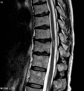
(Space occupying lesion in the spine)


Signs and Symptoms of cauda equina syndrome include the following: 

  • Low back pain
  • Unilateral or bilateral sciatica
  • Saddle and perineal hypoesthesia or anesthesia
  • Bowel and bladder disturbances
  • Lower extremity motor weakness and sensory deficits
  • Reduced or absent lower extremity reflexes. 
Urinary manifestations of cauda equina syndrome include the following:
  • Retention
  • Difficulty initiating micturition
  • Decreased urethral sensation
  • Typically, urinary manifestations begin with urinary retention and are later followed by an overflow urinary incontinence.
The signs and symptoms are unilateral in Cauda Equina syndrome as the lesion is on the already formed nerve roots, in addition the features will always be like lower motor neuron lesion due to the former reason. 

Differential Diagnoses
  • Acute Inflammatory Demyelinating Polyradiculoneuropathy
  • Amyotrophic Lateral Sclerosis in Physical Medicine and Rehabilitation
  • Diabetic Neuropathy
  • Guillain-BarrĂ© Syndrome
  • Multiple Sclerosis
  • Neoplasms, Spinal Cord
  • Neuromuscular and Myopathic Complications of HIV
  • Neurosarcoidosis
  • Spinal Cord Infections
  • Traumatic Peripheral Nerve Lesions

                           
Sagittal MRI of a patient with cauda equina syndrome secondary to a large lumbar disk herniation. (@medscape)


Conus Medularis Syndrome:

Anatomy:
The distal tapering end of the spinal cord is called the conus medullaris. It comprises the coccygeal segments and S2 to S5 and is superiorly continuous with the epiconus (L4 to S1 segments).[4] The filum terminale, a thin, 20 cm-long strand of fibrous tissue, is the continuation of the pia mater of the conus' tapering end downward. Through the coccygeal ligament, this tissue joins the conus to the coccyx, stabilizing the spinal cord. The cauda equina is the inferior continuation of the lumbosacral nerve roots. The conus often ends at the middle third of the L1 vertebra, although it can also be found as low or as high as the middle third of the T11 vertebra.
The conus medullaris give rise to the lumbar sympathetic, sacral somatic and sacral parasympathetic nerves which continue downward within the cauda equina. These nerves have important functions which can be impaired by injury or ischemia


Conus medullaris syndrome is the result of lesions near the spinal L2 level that impact the conus medullaris. Early onset bowel and bladder dysfunction, symmetric lower extremities motor weakness with hyperreflexia, perianal anesthesia, and abruptly intense back pain are among the symptoms.[14]

Etiology:
Known causes of conus medullaris syndrome include 
  • spinal fracture
  • disc herniation 
  • tumors 
  • trauma 
  • epidural abscess 
  • infarction
It is critical to make a clinical distinction between this illness and cauda equina syndrome, which shares similar origins but is distinguished by saddle anesthesia, asymmetric lower extremity motor weakness with hyporeflexia, acute unilateral radicular pain, and late-onset bladder and stool dysfunction.

Generally speaking, cauda equina syndrome mostly causes symptoms of the lower motor neurons as the lesion is on the already formed spinal nerves, whereas conus medullaris syndrome generates aspects of both the upper and lower motor neurons this is because the lesion here is at the level or the spinal cord. So, there will be upper motor neuron signs below the level of the lesion and lower motor neuron signs at the level of the lesion. 

There are many conus medullaris intramedullary lesions. Glial tumors such ependymoma, astrocytoma, and glioblastoma multiforme are examples of neoplastic lesions; non-glial cancers include hemangioblastoma, primitive neuroectodermal tumor, lymphoma, melanoma, and metastasis. Granulomas from diseases like sarcoidosis and tuberculosis, parasitic infections from diseases like cysticercosis and schistosomiasis, demyelination in multiple sclerosis, vascular lesions from diseases like amyloid angiopathy, cavernoma, and vascular malformations, and dysembryogenetic lesions. 


Conus/epiconus infarction in the setting of sickle cell crisis.(White region)
(@Medscape)


Signs and symptoms comparison:


Conus Medullaris Syndrome

Cauda Equina Syndrome

Presentation

Sudden and bilateral

Gradual and unilateral

Reflexes

Knee jerks preserved but ankle jerks affected

Both ankle and knee jerks affected

Radicular pain

Less severe

More severe

Low back pain

More

Less

Sensory symptoms and signs

Numbness tends to be more localized to perianal area; symmetrical and bilateral; sensory dissociation occurs

Numbness tends to be more localized to saddle area; asymmetrical, may be unilateral; no sensory dissociation; loss of sensation in specific dermatomes in lower extremities with numbness and paresthesia; possible numbness in pubic area, including glans penis or clitoris

Motor strength

Typically symmetric, hyperreflexic distal paresis of lower limbs that is less marked; fasciculations may be present

Asymmetric areflexic paraplegia that is more marked; fasciculations rare; atrophy more common

Impotence

Frequent

Less frequent; erectile dysfunction that includes inability to have erection, inability to maintain erection, lack of sensation in pubic area (including glans penis or clitoris), and inability to ejaculate

Sphincter dysfunction

Urinary retention and atonic anal sphincter cause overflow urinary incontinence and fecal incontinence; tend to present early in course of disease

Urinary retention; tends to present late in course of disease


(@Medscape)

Differential Diagnosis:
  • Spinal stenosis
  • Herniation of nucleus pulposus 
  • GBS
  • Peripheral Neuropathy
  • Lumbar Plexopathy
  • Multiple Sclerosis 
  • Vertebral Fracture
  • Polyradiculopathy 
  • Spinal tumor

Comments

Popular posts from this blog

Unraveling the Marvels of the Mind: Your Ultimate Brain Blog!

Unlocking the Mysteries of Lateral Medullary Syndrome: A Journey through Brainstem Disruption